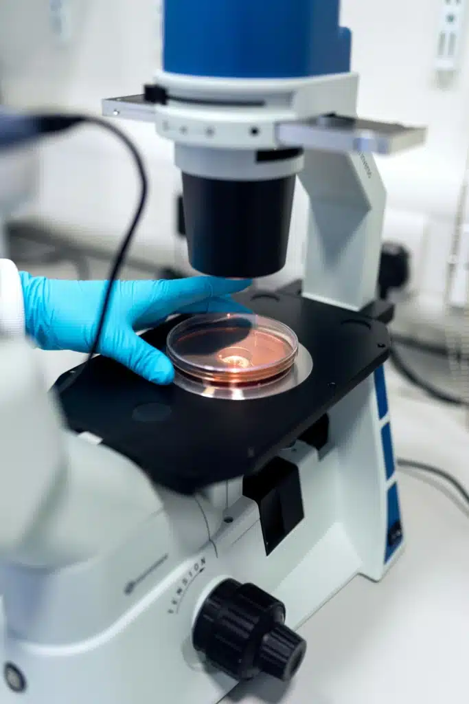
Correlated multimodal imaging (CMI) combines two or more imaging techniques or modalities to gather information about the same specimen, for example, a tumour cell. CMI can provide valuable multidimensional information about a sample and its structure, function, and chemical composition. No single imaging technique can reveal all these details, so advancing CMI is one way to get a better, holistic understanding of a wide range of biomedical processes and diseases.
CMI relies on several different areas of expertise including biologists, physicists, chemists, clinicians, microscopists and computer scientists. As such it requires coordinated activities and knowledge transfer between multiple specialist disciplines. However, no European interdisciplinary network existed in the field until COST Action Correlated Multimodal Imaging in Life Sciences (COMULIS) was established.

Bringing experts together: the COMULIS effect
The Action worked to establish common protocols and quality standards for existing CMI approaches, showcase novel CMI methodologies, and generally raise awareness of CMI to promote interdisciplinary imaging approaches in life sciences and health care.
Professor Andreas Walter of Aalen University in southern Germany was Chair of COMULIS and proposed the Action when he was at Austrian Bioimaging/Vienna BioCenter. “I realised that every field in bioimaging was working on its own,” he explains. “But there was no real cross-talk – no correlated workflows that facilitated going from one imaging technique or modality to another. Each imaging modality or technique can only access certain information and has a limited range of resolution or scale,” Andreas continues. “But if you want to understand something truly holistically you need to gather both structural and molecular data – you need to be able to zoom in and out between all levels. And it is not a trivial issue because different imaging techniques have different sampling techniques, so it is key to be able to reidentify regions of interest from one imaging modality to another.”
“We wanted to make people excited about interdisciplinary approaches and to find a common language”
Prof. Andreas Walter, Chair of COMULIS
The overall aim of the Action was to get clinicians talking to imaging scientists, cell biologists, and structural biologists to really break down barriers and establish communications and resources to ease the combination of different imaging and scientific disciplines.
“We wanted to make people excited about interdisciplinary approaches and to find a common language. We wanted to inspire a new generation of researchers and also tap into the experience of more senior enthusiasts,” says Andreas. “The idea was to push the science forward – to advance the life sciences and have a really positive impact on health care, for example in cancer identification and treatment, by developing imaging approaches further.”


Sustaining the initiative
The valuable work of COMULIS is now going global as ‘COMULISglobe’ with funding from the Chan-Zuckerberg Initiative (CZI): a philanthropic organisation founded by Facebook’s Mark Zuckerberg and his wife Priscilla Chan. “The follow-up grant from the Chan-Zuckerberg Initiative will take our networking and training efforts to a truly global level with participants from Africa, South America and elsewhere. It is potentially a huge network – we already have a mailing list of around 500 people,” says Andreas who is also Chair of COMULISglobe.
COMULIS was also awarded the COST Innovator Grant ‘Faites Vos Jeux!‘ that followed up on one aspect of the Action to automate sample preparation for correlative cryo-microscopy techniques that Andreas was also involved with. “The issue emerged from showcase studies in the original Action that highlighted a bottleneck in workflows for correlated light and electron microscopy (CLEM),” continues Andreas. “This uses two imaging techniques on one sample in an organised way.”
The development of a new sample substrate can both facilitate the use of a single well-characterised sample for both techniques and also introduce automated processes to increase sample throughput. “Our aim was to make life easier for those who do CLEM, which in principle could be any cell or structural biologist in academia, medicine or industry,” concludes Andreas.

